
For decades, spine surgery required large incisions and extended recovery times. It carried a degree of uncertainty that even the most experienced physicians found challenging. The spinal column’s complex structure, surrounded by nerves and delicate tissue, demanded precision that traditional methods could not always achieve. Dr. Larry Davidson, a board-certified neurosurgeon with fellowship training in complex spinal surgery, has focused his practice on approaches that integrate advanced imaging and navigation to improve surgical accuracy and planning. These technologies provide clearer visualization of spinal anatomy and support more consistent outcomes in complex procedures.
Advances in imaging and navigation have transformed the way spinal procedures are performed, enhancing both precision and efficiency in the operating room. Imaging and navigation provide surgeons with detailed views of spinal anatomy and continuous reference during procedures. These tools are used to guide the placement of instruments and implants within defined surgical pathways. Their use has become a central part of procedural planning and intraoperative coordination in many spine practices.
From Static Imaging to Real-Time Visualization
Traditional imaging, including standard X-rays and early CT scans, offered surgeons only static, two-dimensional views of the spinal anatomy. While these methods were critical for diagnosis, they provided limited guidance once surgery began. Surgeons had to rely heavily on experience and anatomical knowledge to navigate deep within the body, where even small inaccuracies could have significant consequences.
Modern imaging technology has changed that. Advanced systems now create high-resolution, three-dimensional maps of the spine in real time. Intraoperative CT scans, fluoroscopic guidance, and computer-assisted navigation provide continuous visual feedback, allowing surgeons to track instruments with millimeter precision.
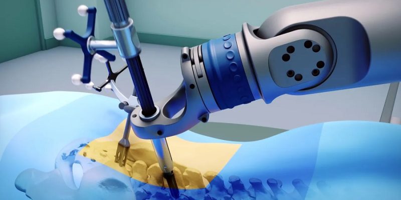
Navigation Systems and Robotic Integration
Intraoperative navigation systems use sensors, cameras, and digital imaging to identify the position of surgical instruments in relation to the patient’s anatomy. This real-time feedback supports precise alignment during the placement of screws, rods, and implants, even within the limited access provided by minimally invasive procedures.
The integration of robotic-assisted systems takes precision a step further. Robots follow pre-planned surgical trajectories generated from patient-specific imaging data, offering stability and control that human hands alone can’t achieve. Surgeons remain fully in command but are supported by tools that increase consistency and minimize risk.
Precision in Patient-Centered Practice
Advancements in imaging have influenced both surgical planning and intraoperative techniques. Improved visualization allows surgeons to operate through smaller access points and follow defined pathways with greater clarity. These approaches are incorporated into care protocols designed to support safety, consistency, and efficiency throughout the surgical process.
Dr. Larry Davidson emphasizes, “Preparing patients for what to expect is just as important as the procedure itself. They come in that morning, get prepped, have the surgery, recover in a dedicated area, and only go home once it is medically safe to do so.” That balance of preparation and precision reflects the broader philosophy behind modern spine care, combining advanced tools with attentive, patient-centered guidance throughout the surgical process.
Supporting Minimally Invasive Techniques
Minimally Invasive Spine Surgery (MISS) relies heavily on advancements in imaging technology. Because surgeons operate through small portals rather than large open incisions, direct visibility is limited. Real-time imaging compensates for this limitation by providing an internal view of the surgical site.
Intraoperative CT, 3D fluoroscopy, and navigation systems are used to guide targeted access to affected areas while preserving surrounding tissue. These imaging tools support the precision required for minimally invasive approaches, which have become a standard option in many spine practices.
Reducing Radiation and Increasing Safety
While imaging is essential to surgical precision, it must also be safe. Modern systems are designed to deliver high-quality images with minimal radiation exposure. Many platforms now use low-dose protocols that protect both patients and surgical teams.
Navigation and robotic systems reduce the number of manual adjustments needed during surgery, which can lessen reliance on repeated imaging. These tools are incorporated to maintain visual consistency and procedural control throughout the operation.
Enhancing Training and Surgical Planning
Imaging technology has also transformed how surgeons train and prepare for operations. Preoperative planning software allows for detailed visualization of spinal anatomy and the simulation of surgical pathways. Surgeons can better anticipate challenges, adjust strategies, and even practice complex cases using virtual models derived from actual patient scans.
This planning extends into the operating room, where real-time imaging confirms that each step of the procedure aligns with the preoperative plan. The integration of these tools has standardized outcomes, improved predictability, and strengthened surgeon confidence across all experience levels.
Improving Outcomes Through Collaboration
Advanced imaging has created new opportunities for collaboration within the surgical team. Anesthesiologists, nurses, and surgical assistants can all see real-time data, enabling smoother coordination during critical moments. Postoperatively, these images guide physical therapy, allowing rehabilitation teams to tailor exercises based on precise structural details.
This continuity between surgery and recovery demonstrates how imaging technology enhances not only the procedure itself, but the entire patient care continuum. Each advancement in visualization brings every member of the team closer to the same goal: safe, efficient, and lasting recovery.
The Next Era: AI and Predictive Imaging
Artificial Intelligence (AI) is rapidly expanding the capabilities of imaging. Algorithms are being developed to detect early signs of degeneration, simulate surgical outcomes, and assist in real-time decision-making during procedures. AI-driven imaging could one day identify patterns invisible to the human eye, further reducing complications and improving efficiency.
These technologies support the surgeon’s expertise, providing additional data and contextual awareness during complex procedures. As imaging and navigation continue to evolve, their role within spine surgery is expected to expand, shaping how future procedures are planned and performed.
Precision That Empowers Patients
The development of imaging and navigation has done more than modernize surgery; it has empowered patients. By reducing uncertainty, improving safety, and accelerating recovery, these innovations have redefined what patients can expect from spine care.
Imaging and navigation technology continue to advance in tandem with clinical judgment and surgical skills. This integration reflects a model of care that values both precision and patient experience, aligning technical progress with the broader goals of safety, coordination, and trust in modern spine surgery.


Comments